When it comes to IVF (In-Vitro Fertilization), every step of the process is crucial in determining the success rate. Among these, oocyte grading—or the process of assessing the quality of eggs—plays a pivotal role in predicting fertilization, embryo development, and ultimately, pregnancy success.
At Reviva IVF, led by Dr. Sandeep Cheema Sohi, one of the most renowned infertility specialists in the region, oocyte grading is performed with precision, care, and expertise. With years of experience in Assisted Reproductive Technology (ART) and a compassionate approach toward patients, Dr. Sohi ensures that even the most challenging cases are given the best chance of success.
What is Oocyte Grading?
Oocyte grading is the evaluation of eggs (oocytes) retrieved during IVF to determine their maturity and quality. Not all eggs retrieved are equal—some may be immature, some abnormally shaped, while others may have the ideal characteristics for fertilization.
Grading helps embryologists decide:
- Which oocytes are most likely to fertilize successfully.
- Which can develop into healthy embryos.
- Which eggs are suitable for freezing (cryopreservation) for future use.
Types of Oocyte Quality Grading
Oocyte grading in IVF is typically based on maturity and morphology (appearance under a microscope):
- Germinal Vesicle (GV) Stage
- Immature eggs with a visible nucleus.
- Not suitable for fertilization.
- Immature eggs with a visible nucleus.
- Metaphase I (MI) Stage
- Eggs that are partly mature.
- May develop further in culture but generally have lower fertilization potential.
- Eggs that are partly mature.
- Metaphase II (MII) Stage
- Fully mature eggs with the highest potential for successful fertilization.
- These are considered the gold standard in oocyte quality grading.
- Fully mature eggs with the highest potential for successful fertilization.
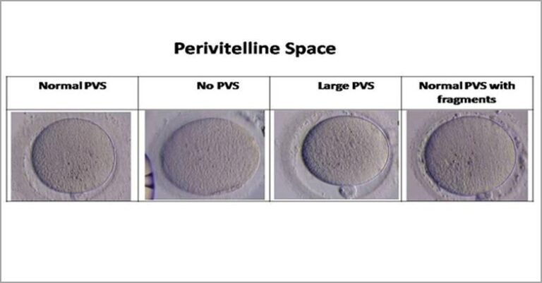
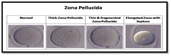
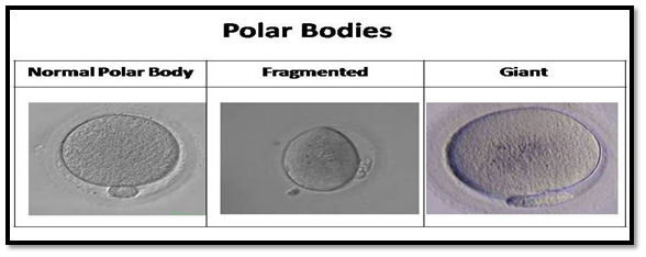
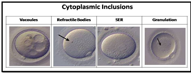
- Abnormal Oocytes
- Eggs with irregular shape, cytoplasmic granularity, or fragmented structures.
- Usually less likely to lead to successful embryo development.
- Eggs with irregular shape, cytoplasmic granularity, or fragmented structures.
Why Oocyte Quality Matters in IVF
High-quality oocytes are directly linked to:
- Better fertilization rates
- Improved embryo quality
- Higher chances of implantation
- Reduced chances of miscarriage
For couples undergoing IVF, understanding that egg quality matters as much as quantity can set realistic expectations and guide personalized treatment strategies.
Factors Affecting Oocyte Quality
Several factors can influence oocyte grading, including:
- Age of the woman: Egg quality declines significantly after the age of 35.
- Hormonal balance: Proper ovarian stimulation is crucial.
- Lifestyle factors: Smoking, stress, alcohol, and poor diet negatively impact egg quality.
- Underlying medical conditions: PCOS, endometriosis, and other reproductive disorders may affect oocyte health.
At Reviva IVF, patients are guided on holistic lifestyle and medical interventions to improve egg quality before treatment.
The Reviva IVF Approach
Dr. Sandeep Cheema Sohi combines advanced technology with personalized care. At Reviva IVF:
- Every oocyte is meticulously graded by expert embryologists.
- The selection process ensures only the best quality oocytes are used for fertilization.
- Patients receive transparent updates on their egg quality grading to stay informed at every stage.
With her empathetic nature and ability to simplify complex medical processes, Dr. Sohi ensures that patients feel reassured and supported throughout their fertility journey.
Final Thoughts
Oocyte grading in IVF is one of the most important steps that determines the likelihood of success. By focusing not just on the number of eggs retrieved but also their quality, Reviva IVF maximizes the chances of achieving healthy pregnancies.
With the expertise of Dr. Sandeep Cheema Sohi and her team, patients are assured of world-class treatment, compassionate care, and a commitment to turning hope into reality.